ASEI Lesson Plan on Biology
Lesson Note on Human circulatory System
Theme: Circulatory System
Topic: Human Circulatory System
Sub Topic: Structure of the Heart
Date: dd/mm/yyyy
Class: S.S.S 2
Duration: 35 Minutes
No of Learners: 30
Learning Objectives:
By the end of the lesson learners should be able to:Define/explain the human circulatory system.
Describe the heart and the structure of the heart.
- the upper right and left chambers are called the auricles also called atrium (plural atria)
- the two lower right and left chamber called the ventricles.
- The heart can contract continuously without fatigue. Therefore can beat for a lifetime without taking a rest.
- Cardiac muscle is also myogenic, which means that its contractions are started by the muscle itself and not by nerves as is the case with other muscle tissue in the body.
Describe the heart valves.
- Two of these valves are called the atrioventricular valves, which allow the blood to flow only from the atria to the ventricles. The one found in the right side of the heart is called the tricuspid valve because it has three flaps. On the left side of the heart is the bicuspid valve. It is also known as the mitral valve.
- The other two valves found in the heart are the semilunar valves. When open, they allow blood to move from the ventricles into the arteries and away from the heart.
List the function of the Heart.
- To receive blood into it at the auricles.
- To send blood out of it to every part of the body at the ventricles.
Describe the circulation of blood in the heart.
The mammalian circulatory system consists of a heart that pumps blood to circulate through a well-defined network of vessels (arteries, veins and capillaries) throughout the body.
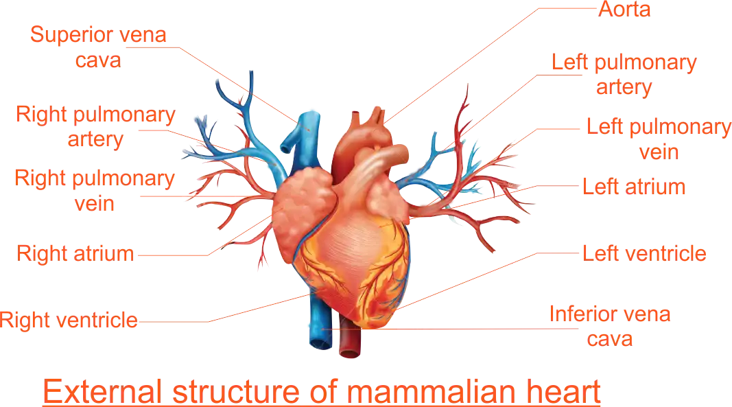
The human heart is located in the thoracic cavity. It lies in-between two lungs. It is a reddish pear-shaped organ that consists of a type of muscle called cardiac muscles. Externally, the heart is covered by a thin membrane called the pericardium. This enclosed fluid, the pericardial fluid, whose function is to greatly reduce friction caused by movements of the heart during pumping.
 External Structure of the Heart
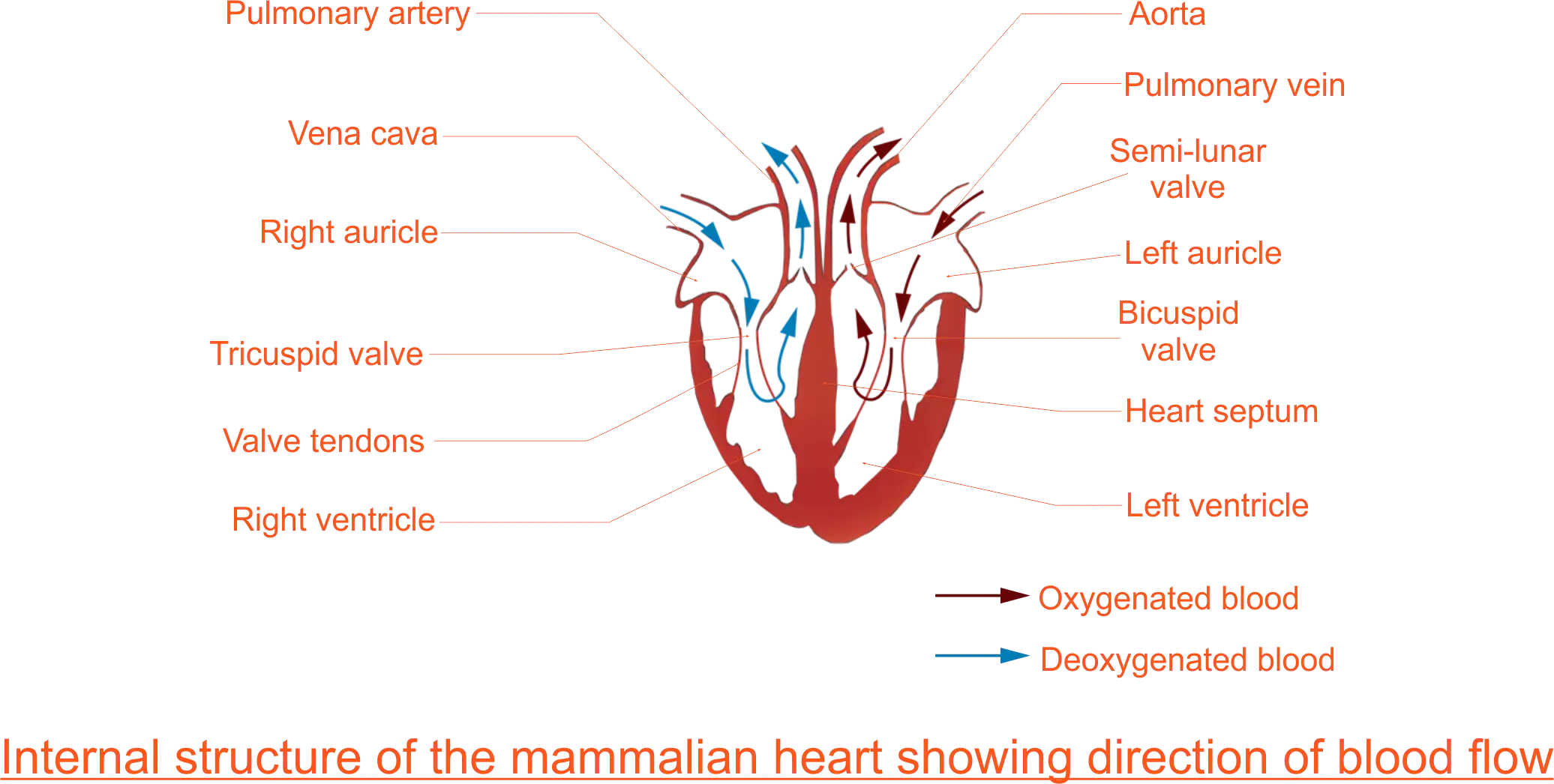
 Internal (Longitudinal) Structure of the Heart
The internal (longitudinal) section of the mammalian heart shows that the heart is divided into two sides, (made up of four distinct compartments or chambers). These are;
The left and the right side are completely separated by a wall called the septum. The septum prevents blood on the right side from mixing with that on the left side. The auricles are smaller, have thin wall and receive blood into the heart which they pump to the ventricles. The ventricles are larger, have thicker walls than the auricles and pump blood out of the heart.
The heart is made of a special muscle called the cardiac muscle. This muscle is special in two ways:
Four flap-like valves control the direction of blood flow inside the heart.
The heart receives blood when its muscles relax. It pumps blood when its muscles contract. These two processes take place in a repeated sequence or cycle known as the heart or cardiac cycle. The cardiac cycle has two alternating phases known as systole and diastole. During systole, the muscles of the heart chambers contract to pump out blood. During diastole, muscles of the heart chamber relax for them to receive blood. The right atrium receives blood coming from the body tissues through the vena cava. This blood has very little oxygen dissolved in it because most of the oxygen has been taken up for respiration by the tissues. It is however rich in carbon dioxide and appears dull red. This blood is described as de-oxygenated blood.
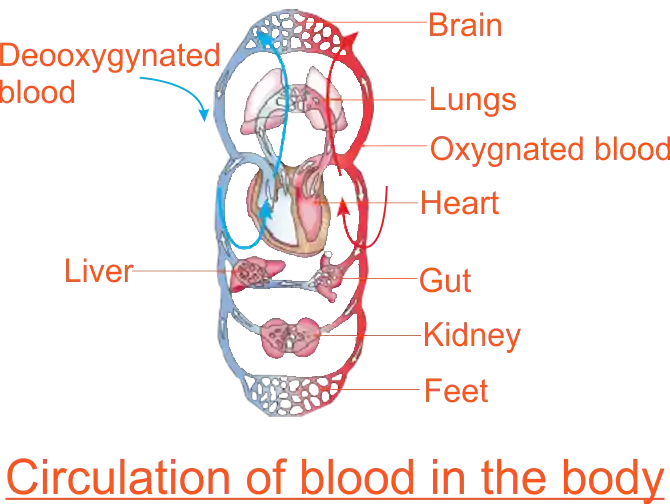
 Circulation of Blood in the Body
The right atrium then pumps the blood into the right ventricle via the tricuspid valve. When full, the right ventricle lets blood into the pulmonary artery. Semi-lunar valves at the opening of this artery prevent backflow into the right ventricle. At the same time, the tricuspid valve prevents any backflow of blood into the right atrium.
The pulmonary artery carries blood to the lungs. In the lungs, the blood picks up oxygen and gives up carbon dioxide. It is now said to be oxygenated and is bright. It goes to the left atrium of the heart via the pulmonary vein.
The left atrium lets blood into the left ventricle via the bicuspid valve. The left ventricle pumps blood to all parts of the body, except the lungs. This blood leaves the left ventricle through the aorta. Semi-lunar valves that open into the aorta prevent the back flow of blood. The left ventricle walls are much thicker than the right ventricle walls to develop a high enough pressure to pump blood to all parts of the body.
Rationale:
The blood makes two types of circulation at the same time. Pulmonary or lung circulation and Body or systemic circulation. Pulmonary circulation takes blood from the heart to the lungs and from the lungs to the heart again. Systemic circulation involves the distribution of blood rich in oxygen to every part of the body except the lungs and the collection of blood whose oxygen has been largely used up back to the heart. The end of the journey is at the capillaries, where oxygen and food in the blood in the blood are discharged to the tissues for cell respiration and food assimilation respectively.
Prerequisite/ Previous knowledge:
- The flow of blood through an opening in the body.
- The human blood and its components.
- Transport of materials.
Learning Materials:
Charts of the circulatory system of a mammal, structure of a sheep or goat’s heart.Reference Materials:
Biology A New Approach For Senior Secondary Schools and Colleges By E. O. Egho,
Modern Biology for SSS By Kucy I. Aunwa et al
Online resources/Internet.
Lesson Development:
| STAGE | TEACHER'S ACTIVITY | LEARNER'S ACTIVITY | LEARNING POINTS |
|---|---|---|---|
| INTRODUCTION full class session (5mins) |
The teacher asks learners to place three fingers firmly on a partner’s wrist, and shift the position of these fingers. teacher asks learners:
|
Learners response to teacher's question.
|
Confirming previous knowledge. |
| STEP 1 Development full class session (5mins) |
The teacher asks learners to explain why blood flow out of an opening/cut on the body whenever there is an opening/cut on any part of the body resulting from the injury. The teacher thereafter asks learners to define/explain the mammalian circulatory system in their own words. |
Learners respond to the teacher's question. The heart acts as the pump for the blood through a network of vessels throughout the body, as the blood circulates throughout the body if there is an opening/cut at any part of the body the blood tends to flow out through that cut/opening. The mammalian circulatory system consists of a heart that pumps blood to circulate through a well-defined network of vessels (arteries, veins and capillaries) throughout the body.
|
Developing the idea of the circulatory system. |
| STEP 2 ACTIVITY 1 (10 minutes) |
The teacher guides learners to form a group to carry out an activity to examine the structure of a sheep or goat’s heart. The teacher provides learners with the following;
|
Activity 1: External Structure of the Heart
 Internal (Longitudinal) Structure of the Heart
The learners form working groups and follow teachers directives.
| Structure of the heart. |
| STEP 3 ACTIVITY 2 (5 minutes) The heart valve. |
The teacher asks the students in groups to study the heart valve closely and outline their features. The teacher gives groups a prompt
|
Learners expected response. The auricles are smaller; they receive blood into the heart through five main blood vessels.
The ventricles are larger; they receive blood through the openings from the auricles guarded by flap-like valves. These are:
|
Describing the heart valves. |
| STEP 4 ACTIVITY 3 (5 minutes) Circulation of blood in the heart. |
The teacher provides learners charts. The teacher guide learners to;
|
Activity 3: Circulation of Blood in the Body
Learners expected response.
The Learners list the advantages of a double circulatory system over a single circulatory system. |
Circulation of blood in the heart. |
| EVALUATION (5 minutes) |
The teacher evaluates the lesson by posing questions;
|
|
Asking the learners questions to assess the achievement of the set objectives. |
| CONCLUSION 5 mins |
The teacher consolidates the main points and corrects any misconceptions. The heart tissue itself receives food nutrients and oxygen via a vessel known as the coronary artery which branches from the aorta and spreads through the heart muscle. The auricles and ventricles have the power to contract. Whenever that happens, blood is pumped away from them. First, the auricles contract and the blood in them is pressed into the ventricles. The blood passes through the valves. As the ventricles fill with the blood, the valve shut up, to prevent the blood from rushing back into the auricles. Next, the ventricles contract and the blood is pumped out of the heart. From the right ventricle, the blood is pumped into the two lungs by way of the pulmonary arteries. From the left ventricles, the blood with a very great pressure is forced into the aorta and thence to the different arteries of the body. When the ventricles are contracting, the auricles are relaxed and it is at this time that the auricles fill with blood. The pumping action of the heart is referred to as the heartbeats. The origin of the heartbeat is in the right auricle at a spot called the sinoatrial node ( or pacemaker). The contraction phase of the heart (ventricles) is called systole while the relaxation period which follows contraction during which time the two auricles of the heart expand and fill with blood is called diastole. heartbeats per minute varies with different mammals and usually under different conditions. The number of beats for an adult man when at rest is 72, for a rat it is 200, for the rabbit is 100 and for an elephant, it is just 12. The instrument used to listen to the beating of the heart is called a stethoscope. |
Learners consolidate the key areas of drawing and labelling plan diagrams. | The main points are no shading, continuous lines, use of sharp pencil and to draw what is observed. |
|
|
|||
| ASSIGNMENT |
|
Learners answer other questions | Improving their level of understanding |